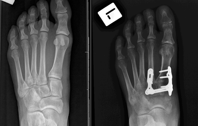
Friday, September 1, 2017

Six months post fusion

I can hardly believe it has been six months since my fusion surgery of my first metatarsal, first cuneiform, and medial cuneiform. So far, it has been a journey with many ups and downs. Fortunately, there have been fewer downs than ups, which means the net effect has been progress. I am happy to say that my foot has not felt this good since before my whole Lisfranc fiasco - starting 3 years ago almost down to the day this entry is published.

I began a walking regimen, starting at 3.5 months post surgery (2.5 months ago, June 2017). My main goal was to first and foremost rehab my gait and learn to walk correctly. However, just as importantly, I wanted to slowly grease the gears of my muscles and tendons in my legs, ankles, and feet that hadn't been properly utilized in years. I think the best analogy to me getting moving again after my surgery would be like starting a 1960 Ford pickup that had been laying out in the yard for 30 years without being driven. Initially, with a new battery and an oil change, the truck may start up after a few cranks. After getting it started, you may be able to drive it around the block slowly with no problems. But once you take the truck out on the main road and drive it faster and for longer distances, all kinds of problems pop up: oil leaks, stiff suspension, the radiator needs to be replaced, the transmission needs a flush, the belt fan breaks, the brakes are shot, etc... If I haven't lost you, just replace all the problems that arise when driving an old truck with the maladies that I experienced when I started walking again. 

Aches, pains, and swelling - not limited to my feet - all graced me with their presence in strange and often confusing ways. One week it was a soreness/pain along my ankle and the next week it was a soreness/pain on the side of my arch. At some points, I thought I had hurt myself by pushing it too much only to be surprised the next day when the pain or soreness had disappeared. I have struggled with plantar fasciitis and posterior tibial tendonitis on and off for the past 2.5 months. Right now I have a nagging case of tendonitis on my right leg - not even the side that I had surgery on 6 months ago - that has popped up for reasons I can't explain. I am not even partaking in high impact sports or running of any sort... So goes the mystery of the adjusting body.

Nearly 3 months into my walking regimen I have worked my way to walking 2 miles 3-4 times a week. To give some perspective, when I started I walked 1/4 miles at a crawl. I slowly worked on more distance week by week. Additionally, I worked on my stride and walking speed. Now I can walk just as fast a normal person. Between walking, I also did other strengthening exercises to rehab my legs, feet, and overall body including swimming, weight training, and stationary biking.

I have recently been doing a little hiking. Nothing too impressive but nonetheless something I haven't been able to do in years. I first started hiking a one-mile path up a small mountain close to home, and then I progressed to a two-mile hike up a more demanding mountain in the Blue Ridge. Besides the pesky posterior tendonitis in my right leg, my feet felt fine and the hike was good. Below is a picture I took from the top of the mountain.


Wednesday, June 7, 2017

Threshold

It has been 14 weeks since my fusion surgery of my first metatarsal-cuneiform and first cuneiform-second cuneiform in my left foot. My last post detailed my progress weight bearing in a boot. In total, I spent 6 weeks in a boot. The first 2-3 weeks of which were filled with ups and downs related to pain, soreness, and stiffness in my foot and ankle. However, I was usually able to abate any pain or swelling with icing and rest. After about 3 weeks in the boot, most of the kinks were worked out and my foot and ankle were fine.

Having gone through this charade before, I was relatively happy with the progress of my foot while in a boot. I have slowly regained mobility and strength in my foot and ankle, which was nearly nil directly after my NWB cast was removed. The new found mobility that the boot afforded me was better than the best Christmas gift I ever received. My mobility gave me the freedom and independence that had been shying away from me for the 5 months I have been dealing with this particular foot problem. Because of this, my general mood and motivation have improved significantly.
Oblique X-ray showing my hardware and signs of fusion
of the 1st TMT joint.
Now after 6 weeks in a boot, it is finally time to approach the threshold moment in recovery - shed the moon shoe to make one more small step towards healing. This brings us up to speed with my progress as of today. This morning I went to the doctor and was cleared - after standing weight-bearing X-rays were analyzed - to transition to walking in a normal shoe. As in any step in this recovery, I will have to work out a few kinks before this next stage of freedom is complete. I imagine I will experience some pain, swelling, and soreness in the weeks to come as I wean myself off of the boot and mature to a shoe. Most importantly, I will have to listen to my body, more specifically my foot, in this next stage. As long as I feel I am making progress, I will be happy.

My cat sniffing my newly freed foot. I would guess it didn't
smell like roses.
Today marks 6 months since I last stood on the ground with both feet sans boot, crutches, or knee scooter. The mere fact that I can possibly walk unencumbered with my two feet makes me extremely happy but also anxious. Since I have gone through this terribleness - in some regard or another - four times previous, I cannot help but have thoughts of my foot somehow becoming hurt again. I do my best to assuage and process these feelings so that I am able to stay positive and look forward. For any of you veteran Lisfrancer's or the unfortunate newly indicted members that are reading this, you probably have some sense of this. It has been a long and arduous road and I am still on it. Patience is key. But I sincerely hope I am leaving behind an unfortunate period and moving on to better times - at least in regard to my feet.


For anyone with questions regarding Lisfranc injuries, treatment options, and recovery please feel free to ask any question you have. I don't claim to be all knowing about this type of injury - and as I have stated many times previous every Lisfranc injury is different - but I have a large amount of knowledge through experience and research on the subject. Today marks 3 years that I have been dealing with this injury. As far as doctors, treatments, and recoveries I have been through the gambit. I would love to use these otherwise useless credentials to help others because I know how frightening it is to not know. I can't put these things on my resume so I might as well use them somehow...

Pictures of animals always make things better.
Here my cat looks like a pear.

Wednesday, May 10, 2017

The Afterburners

It has been 10 weeks post fusion surgery on my left foot. I have spent the last two weeks transitioning from non-weight bearing in a hard cast to partial weight bearing with a boot. Honestly, it has been a slow go, but I have been making progress, which is all I am worried about. At the moment, I have graduated from partial weight bearing with two crutches and a boot to partial weight bearing and one crutch and a boot. Little by little I have put more pressure on my foot over the course of 14 days. This has ensured that I not only decrease the risk of injury, but I also build up stamina and strength in my foot. In effect, this type of transition lowers the amount of pain I experience each day. My foot is fairly swollen and sometimes sore or painful in the evening or after a day of greater exertion on my foot. However, the swelling and pain have been decreasing quite consistently with each passing day (hopefully I don't jinx myself).  
Dorsal X-ray of left foot 8
weeks post fusion surgery.

The biggest fear I had through this process is that my joints would not fuse properly. Nevertheless, I have been reassured by my doctor that the X-rays show good signs of a proper fusion. The progression to "walking" is a big step in any recovery of this nature. My spirits are definitely higher with my two legged - although still somewhat constrained - freedom. Being that this is my 5th surgery on my feet in less that a year and a half, I really hope that it was my last. As anyone who has gone through a Lisfranc injury and subsequent surgery can attest, it is overwhelming and painful - both physically and mentally. Psychological integrity is imperative to push through these types of injuries and recovery. In truth, mine has been tested beyond anything I ever imagined and worn down to the point of being nearly broken. I hope I emerge from this in a healthy state. 

When I first found out I had a Lisfranc injury, I scoured the internet to find others with similar injuries. I wanted to learn my options as well as the outcomes. For those who are now just discovering they have a Lisfranc injury and all the horrible anxiety that comes with it, I hope that you find this blog helpful. I tell almost everyone that has a Lisfranc injury and is looking for answers that every Lisfranc injury and recovery is different. Do not go looking for specifics. It should never be expected that one person's experiences - whether positive or negative - are transferrable to your own situation. But, it is useful to be knowledgeable about Lisfranc injuries, how others were treated, and the possible outcomes of each type of treatment. I hope that one day I will be walking around on two feet again in relative painlessness. Until then, I have to put on the afterburners and keep on truckin'.

Science has proven that looking at pictures of dogs and cats can lower one's stress levels and blood pressure... So in closing, here are some pictures of my cat:


Thursday, April 27, 2017

Out of the fire and into the frying pan

When I was in the cast. Foot is resting on my knee scooter
(best investment I made).
     It has been about 8 weeks since I last posted. At the time, I had just undergone fusion surgery on my left foot. Two weeks post-op my surgical splint was removed, X-rays were taken to ensure hardware was in place, and sutures were removed from the surgical incision. X-rays and incision all looked good. After the splint was removed, I was placed in a non-weight bearing hard cast, which I wore for 6 weeks. Life in a cast was frustrating, boring, and slow. I had a knee scooter for getting around the house easily. I learned from previous surgeries on my foot that freeing up my hands - which crutches do not allow - with the knee scooter enabled me to be considerably more independent. The only times I used crutches were if I went out of the house with my family or to see a friend. 

     Now almost two months after my operation, I have finally been removed from my non-weight bearing duties in the hard cast, and I have been transferred into a walking boot. This doesn't necessarily mean that life is any easier now. Putting pressure on my foot for the first time - even in a boot - is a painful process. In addition, I have to learn how to walk again. I am actually slower at getting around now than when I was in a cast. Nonetheless, I have been happy to walk (although very slowly) on two-ish feet. The transfer into a boot is a slow process, and I know it will take days if not a week or two for me to be relatively comfortable walking in the boot. I got to get the gears greased and cranking again.
The cast is off and into the boot. Had to include a picture of
the incision.
In the meantime, my cat (Luna) has been keeping me company and I have added a few more books to my completed reading list.

1. Unbroken: A World War II Story of Survival, Resilience, and Redemption - Laura Hillenbrand
2. Stiff: The Curious Lives of Human Cadavers: Mary Roach
3. Tuesdays With Morrie: An Old Man, a young Man, and Life's Greatest Lesson
4. Hiroshima: John Hershey

Luna in loaf mode


Wednesday, March 8, 2017

Lucky number 5

I had surgery eight days ago to fuse my first metatarsal, intermediate, and medial cuneiform. I now have six screws and a metal plate in my left foot. Coincidently, the surgery was a year to the day after my hardware removal surgery on the same foot - originally had ORIF operation on left foot in October 2015 and hardware removal February 28, 2016. 

By now I should be used to these types of surgeries, - this was my 5th Lisfranc related surgery on my feet in the past year and a half - but the pain, nausea, and dizziness never get any better. The pain and swelling from the surgery were the greatest in the first few days. Initially, it felt as if a sledge hammer was pounding a railroad spike into my midfoot. This pain tapered off around day three, which is also when I began weening off my pain medication. Personally, coming off the pain medication was nearly as awful as the initial pain from surgery. Nausea and dizziness were my two least favorite friends that didn't leave my side for days following my surgery. My head spun for three or four days after I stopped taking the pain meds. Eventually, my body detoxed itself and I have felt better since. 

I have been religiously keeping my foot elevated to reduce swelling and pain. Each day my foot feels a bit better. It is nice to feel healing progress. I take a calcium supplement every day to aid a healthy fusion of the joints. My surgeon says I have to be non-weight bearing in a cast for 8 weeks. I go to the doctor in one week to take off my splint and take out the sutures from the surgical incision. After that, I will be put into a medical boot instead of a hard cast. 

Sadly, I have been through this process numerous times now. I know the steps by heart. Thinking about it doesn't sadden me until I take a moment to let it sink in that this routine has encompassed my life for over a year. I can't remember what life is like not in a continuous state of surgical recovery. Hopefully this is lucky number 5...

In other thoughts... I have compiled the list of books I have read over the past year. I am proud to say that I have nearly read more books in a year than I have read the entire preceding 25 years of my life. Since surgery, I have already read three books. It took me until I was no longer in school to finally realize the importance and joy of reading. I hope all my past teachers would be proud nonetheless. Better late than never.
  1. The Memoirs of Sherlock Holmes - Sir Arthur Conan Doyle
  2. Walk Across America - Peter Jenkins
  3. A Walk in the Woods - Bill Bryson
  4. Lost on the Appalachian Trail - Kyle Rohrig
  5. The Appalachian Trail: a journey of discovery - Jan D. Curran
  6. Hiking Through: One Man's Journey to Peace and Freedom - Paul Stutzman
  7. Killing Kennedy: The End of Camelot - Bill O'Rilley and Martin Dugard
  8. Lord of the Flies - William Golding
  9. Stay - Allie Larkin
  10. Life of Pi - Yann Martel
  11. Under the Banner of Heaven: A Story of Violent Faith - Jon Krakauer
  12. Into Thin Air: A Personal Account of the Mt. Everest Disaster - Jon Krakauer
  13. Where Men Win Glory: The Odyssey of Pat Tillman - Jon Krakauer
  14. Into the Wild - Jon Krakauer
  15. The Lost City of Z: A Tale of Deadly Obsession in the Amazon - David Grann
  16. Eat and Run - Scott Jurek
  17. Finding Ultra - Rich Roll
  18. Dividing the Great - John Metcalfe
  19. The River of Dought: Theodore Roosevelt's Darkest Journey - Candice Millard
  20. Isaac's Storm: A Man, a Time, and the Deadliest Hurricane - Erik Larson 
  21. Dead Wake: The Last Crossing of the Lusitania - Erik Larson
  22. Devil in the White City: Murder, Magic, and Madness at a Fair that Change America - Erik Larson
  23. In the Garden of Beasts: Love, Terror, and an American Family in Hitler's Berlin - Erik Larson
  24. Chelsea Chelsea Bang Bang - Chelsea Handler
  25. Is Everyone Hanging Out Without Me? - Mindy Kailing
  26. Not That Kind of Girl: A Young Woman Tells You What She's Learned - Lena Dunham
  27. Eat Sleep Ride - Paul Howard
  28. Book 1: Harry Potter and the Sorcerer's Stone - J.K. Rowling
  29. Book 2: Harry Potter and the Chamber of Secrets - J.K. Rowling
  30. Book 3: Harry Potter and the Prisoner of Azkaban - J.K. Rowling
  31. Book 4: Harry Potter and the Goblet of Fire - J.K. Rowling
  32. Book 5: Harry Potter and the Order of the Phoenix - J.K. Rowling
  33. Book 6: Harry Potter and the Half-Blood Prince - J.K. Rowling
  34. Book 7: Harry Potter and the Deathly Hallows - J.K. Rowling
  35. Bury My Heart at Wounded Knee - Dee Brown
  36. Crazy for the Storm: A Memoir of Survival - Norman Ollestad
  37. Skywalker: Close Encounters on the Appalachian Trail - Bill Walker
  38. AWOL on the Appalachian Trail - David Miller
  39. Wild - Sheryl Strayed
  40. The Oregon Trail: A New American Journey - Rinker Buck

Saturday, February 11, 2017

The Lisfranc injury: Symptoms, Diagnosis, and Treatment Options

Background and Anatomy

The Lisfranc joint complex is composed of the five tarsometatarsal (TMT) joints, which are important for the articulation between the forefoot and midfoot. This anatomic region is named after the 18-19th-century French surgeon Jacques Lisfranc de St. Martin. He was a field surgeon for Napoleon's army serving on the Russian front and developed a new amputation technique across the five TMT joints as a treatment for soldiers with gangrene or crush injuries to the midfoot. Now known as the Lisfranc joint, the term is used today to describe a variety of traumatic injuries to the TMT joints in the midfoot.

Image result for lisfranc injury
Anatomy of Lisfranc joint complex
The TMT joints--also known as the Lisfranc joints--involves the first, second, and third cuneiform and metatarsal bones, and the cuboid bone. The Lisfranc joint can be divided longitudinally into three columns, which include:

  • Medial column, or first ray, consisting of the medial cuneiform and first metatarsal
  • Middle column, consisting of the second and third metatarsals and cuneiforms
  • Lateral column, consisting of the fourth and fifth metatarsals and cuboid
Crucial plantar and dorsal interosseous ligaments link and stabilize the Lisfranc joint complex. The dorsal ligaments are weaker than the plantar ligaments which can explain the majority of Lisfranc injuries involving dorsal dislocations. The first of the dorsal ligaments connects the first (medial) cuneiform and base of the second metatarsal. The second connects the second (intermediate cuneiform) to the base of the second metatarsal. Lastly, the third stabilizing interosseous ligament connects the third cuneiform with the third metatarsal. The proper stabilization/maintenance of these ligaments and bones within the Lisfranc joint complex is crucial for proper function of the foot upon weight bearing. Any rupture of the interosseous ligaments or fracture-dislocation of the joint complex can destabilize the midfoot.

Example of the common location of a Lisfranc fracture and ligament tear (left) and the mechanism of the Lisfranc injury with displacement of the second metatarsal (right).

Mechanism of Injury by Percentage of Occurrence
Most common mechanisms of Lisfranc
injury from Shahin Sheibani-Rad, MD,
MS et al. 2012
Lisfranc injuries to the midfoot are a rare (1 in 55000 persons) but debilitating injury. They occur when large shearing or crushing forces affect the TMT/Lisfranc joint complex in the foot. When large forces are stressed across these ligaments they can be sprained, torn, or completely ruptured. This leads to instability in the midfoot upon weight bearing. Common symptoms of the Lisfranc injury include swelling and discoloration of the dorsal and plantar aspects of the midfoot, pain upon palpation of the midfoot, pain instigated by dorsal or plantar flexion of the foot, and pain present when pronating or supinating the foot. Injury to the Lisfranc joint complex is common in high energy trauma such as in motor vehicle crashes or falls from height. There has also been an increased uptake in Lisfranc injuries in athletes such as football and soccer players, gymnasts, and horseback riders. Regardless of the mechanism of injury, a TMT dislocation or fracture-dislocation leading to midfoot instability or malalignment is a serious injury and should be treated as soon as possible. 

Diagnosis

Image result for lisfranc injury
Weight-bearing X-rays showing an example of a normal
Lisfranc joint (left) and a dislocated Lisfranc joint (right).
Delayed diagnosis and treatment of Lisfranc injuries can lead to further joint damage, deformity of the foot, and chronic pain. Successful recoveries and a good prognosis are heavily dependent on a timely diagnosis and proper course of treatment. However, Lisfranc injuries, particularly subtle ones, are commonly missed or misdiagnosed. Imaging tests such as X-ray and MRI are commonly utilized as a diagnostic tool for Lisfranc. Weight-bearing radiographs are standard for determining the degree of instability or displacement between the metatarsals and cuneiforms in the midfoot. MRI's can also be helpful for indicating ligamentous and intraarticular damage or bone fractures that often do not appear on an X-ray. Nevertheless, it is common for subtle displacement, malalignment, or fractures to go unnoticed in X-ray and even MRI imaging. In these cases, manipulating/stressing the midfoot by abducting the first ray (first metatarsal and first cuneiform) from the second ray (second metatarsal and second cuneiform) can provide information on the instability or presence of pain in the midfoot. 
Weight-bearing X-rays of obvious and subtle diastasis/dislocation
of the Lisfranc joint.

Treatments

Image result for degree of lisfranc injury
Different degrees of a Lisfranc injury. Stage I being a spain, Stage II being a
ruptured ligament with diastasis in the Lisfranc joint, and Stage III being a
ruptured ligament with significant diastasis and loss of arch height.
Every Lisfranc injury is unique so it is difficult to determine when to surgically intervene or treat non-operatively with conservative measures. The course of treatment is usually based on the severity of instability/displacement in the midfoot upon stress or weight bearing and the type of Lisfranc injury. The degree of midfoot instability is categorized by the amount of diastasis in the Lisfranc joint:

  • Stage I (a sprain <2 mm)
  • Stage II (2-5 mm) 
  • Stage III (>5 mm)

The type of Lisfranc injury can be classified into purely ligamentous and combined ligamentous and osseous (fracture-dislocations). Conservative treatment is usually advised for Stage I subtle, ligamentous non-displaced Lisfranc injuries. It involves non-weight bearing in a cast or medical boot for 6-8 weeks. Most of the literature agrees that when Lisfranc injuries, whether it is ligamentous or fracture-dislocations, have larger displacement and greater instability (Stage II and IIII) they should be treated operatively. 

The two main types of surgical treatment include open reduction and internal fixation (ORIF) and primary arthrodesis (fusion). ORIF involves the rigid fixation and anatomic reduction of the Lisfranc joint with hardware such as titanium screws, bridging plates, K-wires, or even "synthetic ligaments" (a.k.a. mini-tightrope/suture button). The hardware acts to stabilize the joint complex and allow the ligaments and soft tissue to heal. Often, the hardware is left in the foot for 4-6 months until the stabilization of the midfoot is sufficient and then it is removed. In some cases, the hardware remains permanently unless it causes pain to the patient or breaks. 

Primary arthrodesis surgically fuses the afflicted Lisfranc joints to stabilize the midfoot. The bones in the midfoot are fused by first debriding the injured joints then grafting some of the patients own bone (often from the fracture site, heel, or hip) into the joint space. The joint is closed and stabilized with titanium screws or plate. The number of bones/joints fused relies on the severity and displacement of the injury. If the extent of intraarticular and ligamentous damage is severe in a joint, that joint is usually fused. 

Image result for orif lisfranc
X-ray images of foot with Lisfranc injury pre-op (left) and post-op after ORIF with
metal plates and screws (right).

The preferred type of surgical intervention for Lisfranc injuries has been a topic of debate. There are many factors which are taken into account when deciding on ORIF or primary arthrodesis. Most of the studies advocate for ORIF in:

  • Less severe injuries
  • Non-chronic injuries 
  • Patients who are young 
  • Subtle injuries with slight displacement
  • Injuries with little to no articular damage within the joint 
  • Injuries in high-level athletes

Indications for primary arthrodesis are as follows:

  • Severe Lisfranc fracture-dislocations
  • Injuries in older patients
  • Failed previous ORIF surgery or any Lisfranc injury with persistent pain
  • Presentation of severe osteoarthritis in the joint complex
  • For a long term misdiagnosed or chronic injury

ORIF

Most of the current literature supports ORIF as the primary surgical treatment for Lisfranc injuries that are not severe. It has been shown that ORIF can provide a successful recovery, return to activity, and a good functional outcome following the indications for ORIF of the Lisfranc Joints. Depending on the type of Lisfranc injury, screws, plates, K-wires, or a mini-tightrope can be used to fixate the joint. Although past surgical treatments have mainly involved screws, plates, and K-wires, recent studies have shown that for some types of Lisfranc injuries a mini-tightrope/suture button is useful and has excellent outcomes. In particular, the use of a mini-tightrope/suture button has proven successful in athletes with subtle Lisfranc injuries. 

There is often no need to remove hardware after healing is completed (the mini-tightrope is always permanent) unless there is a hardware failure of the patient experiences pain from prominent screws. However, ORIF can frequently lead to the formation of post-traumatic arthritis within the Lisfranc joint, which can prove to be debilitating and require secondary arthrodesis surgery. Arthritis is usually created from articular damage from the initial injury or from the placement of stabilizing screws through the articular surface in the Lisfranc joint. In order to circumvent the latter of these causes, it is common for surgeons to utilize titanium plates to bridge and stabilize the joints without disrupting the joint surfaces. Nonetheless, some studies have noted that greater than 50% of ORIF patients go on to have arthritis within the affected Lisfranc joint(s). There is also evidence that in purely ligamentous injuries treated with ORIF the healing of the ligaments did not provide sufficient strength to maintain the stability of the midfoot. The compromised integrity of the Lisfranc joint can lead to pain, deformity of the foot, or post-traumatic arthritis. In such cases, a patient with a failed ORIF usually requires secondary surgery involving arthrodesis of the affected joints.

Arthrodesis

In the past, it was thought arthrodesis was done as a last resort for a failed ORIF or very severe fracture-dislocation. However, in more recent studies there has been some controversy as to whether it is better to treat primarily ligamentous Lisfranc injuries with ORIF or arthrodesis. A number of studies provide evidence that primary arthrodesis could provide a better outcome than ORIF in purely ligamentous injuries. One study found that a sample of patients that underwent primary arthrodesis and ORIF for purely ligamentous injuries returned to 92% and 65% of their pre-injury activity level, respectively. Another study compared the postoperative pain levels in a group of arthrodesis and ORIF patients. They found that the arthrodesis group had a mean AOFAS post-surgical pain score (out of 100) of 86.9 compared to 57.1 in the ORIF group. However, other studies have noted that they have found no significant difference between the clinical outcomes of patients who underwent ORIF and primary arthrodesis. 

There is agreement that a primary arthrodesis can potentially prevent the development of a painful, deformed foot, in the case of a failed ORIF, and the need for future surgery. Primary arthrodesis has been found to result in statistically fewer follow-up surgeries compared to that of ORIF if hardware removal is performed. In addition, patients who have been treated with arthrodesis for primarily ligamentous Lisfranc injuries have been shown to function as well as those treated with ORIF. Nonetheless, arthrodesis or partial arthrodesis of the Lisfranc joint has its drawbacks. When the medial or middle column of the midfoot is fused the Lisfranc joint becomes rigid. Therefore, much of the energy that was once absorbed by the Lisfranc joint upon weight-bearing is distributed to adjacent, unfused joints in the foot. Over time, this can lead to the development of arthritis in the adjoining joints or the increased risk of stress fractures. Arthrodesis of the medial column also inhibits mobility in the midfoot, which is thought to be necessary for proper function of the foot when running. For this reason, arthrodesis is usually not the preferred treatment for patients who want to return to high-level athletics. Yet, there have been studies that found the return to athletics in a group of young patients with primary arthrodesis was very high.

Adapted from MacMahon et al. 2016. Pre- and postoperative activity levels of patients in a
 study who received primary arthrodesis. 

Recovery

Recovery from a Lisfranc injury, whether by means of conservative or operative treatment, is a slow process. For the Stage I non-displaced Lisfranc injury, conservative treatment usually involves non-weight bearing in a cast or medical boot for 6-8 weeks. Then the patient is transferred into a partial weight bearing walking boot for another 2 weeks and then full weight bearing in the boot for 4-6 weeks. Physical therapy is advised after the first 6-8 weeks and a transfer to stiff soled shoes and orthotics is recommended after 6-8 weeks in the boot. In total, the recovery period last approximately 3-4 months. 

The recovery period for operative treatment of Lisfranc injuries is similar to that of conservative approaches. Regardless of the type of surgery (ORIF or arthrodesis), the patient is put in a non-weight bearing splint for 10-14 days at which point the split is removed and the stitches are removed from the surgical incision. Once the stitches are removed, a hard cast or air cast is placed on the foot. The cast is removed after 4-6 weeks and weight bearing X-rays are taken to ensure hardware has not moved or become broken. The patient is then placed in a partial weight-bearing walking boot for 7-14 days. Full weight bearing is permitted with the boot after this period and is continued for another 4-6 weeks. More weight bearing X-rays are taken once the boot is removed, and the patient is then transferred into a supportive, stiff-soled shoe with orthotics. 

In both the conservative and surgical treatments, physical therapy can be initiated after the transfer to partial weight bearing in a boot. Therapy usually focuses on regaining proper gait by strengthening the leg, calf, and ankle of the affected leg. The intrinsic muscles of the foot are also worked to strengthen and regain function.

Conclusion

The Lisfranc joint complex provides stabilization to the midfoot and arch upon weight bearing and normal gait. Any tear, rupture, or fracture to the Lisfranc joint can compromise the integrity of the midfoot leading to malalignment and diastasis within the Lisfranc joint. The common mechanisms of injury to the Lisfranc joint include high-energy injuries such as motor vehicle accidents or falls from heights and low-energy injuries that often occur in athletics when the foot is twisted or bent under pressure. 

Timely diagnosis and treatment of a Lisfranc injury are imperative for a good recovery. Weight-bearing X-rays are standard for determining if a Lisfranc injury exists. The weight-bearing X-rays can indicate diastasis or malalignment in the midfoot that is associated with Lisfranc fracture-dislocations. MRI imaging can also be useful to determine the amount of damage to ligaments, soft tissue, and joints that are not visible on X-ray. However, the complexity of the Lisfranc injury makes it easy to misdiagnose or miss altogether. Subtle Lisfranc injuries are particularly difficult to diagnose.

Treatment of Lisfranc injuries is related to the degree of instability/separation in the Lisfranc joint complex. Subtle non-displaced (usually Lisfranc sprains) do well with conservative treatment. More severely displaced injuries require operative treatment to realign/stabilize the joint and heal ligaments. The two main surgical treatments for Lisfranc are ORIF and primary arthrodesis. The severity/type of the injury, the amount of joint damage, and age are the main factors when deciding whether to proceed with ORIF and arthrodesis. ORIF and arthrodesis have both proven to provide good functional outcomes and a relatively high return to activity. Complications of ORIF include the development of arthritis in the Lisfranc joint and possible failure to provide adequate stabilization to the midfoot. Complications of arthrodesis include the development of arthritis in joints adjacent to the fused joints, increased risk of fractures, and a smaller range of motion/function of the midfoot.


References and Resources

Brin, Y. S., Nyska, M., & Kish, B. (2010). Lisfranc injury repair with the TightRope™ device: a short-term case series. Foot & ankle international31(7), 624-627.

Charlton, T., Boe, C., & Thordarson, D. B. (2015). Suture button fixation treatment of chronic Lisfranc injury in professional dancers and high-level athletes. Journal of Dance Medicine & Science19(4), 135-139.

Cochran, G., Renninger, C., Tompane, T., Bellamy, J., & Kuhn, K. (2016). Treatment of Low Energy Lisfranc Joint Injuries in a Young Athletic Population Primary Arthrodesis Compared with Open Reduction and Internal Fixation. Orthopaedic Journal of Sports Medicine4(7 suppl4), 2325967116S00172.

Faciszewski, T., Burks, R. T., & Manaster, B. J. (1990). Subtle injuries of the Lisfranc joint. The Journal of Bone & Joint Surgery72(10), 1519-1522.

Hawkes, N. C., Flemming, D. J., & Ho, V. B. (2007). Subtle Lisfranc injury: low energy midfoot sprain. Mil Med172(9), 12-3.

Lewis Jr, J. S., & Anderson, R. B. (2016). Lisfranc Injuries in the Athlete. Foot & Ankle International37(12), 1374-1380.

MacMahon, A., Kim, P., Levine, D. S., Burket, J., Roberts, M. M., Drakos, M. C., ... & Ellis, S. J. (2016). Return to sports and physical activities after primary partial arthrodesis for Lisfranc injuries in young patients. Foot & ankle international37(4), 355-362.

Mulier, T., Reynders, P., Dereymaeker, G., & Broos, P. (2002). Severe Lisfrancs injuries: primary arthrodesis or ORIF?. Foot & ankle international23(10), 902-905.

Panagakos, P., Patel, K., & Gonzalez, C. N. (2012). Lisfranc arthrodesis. Clinics in podiatric medicine and surgery29(1), 51-66.

Panchbhavi, V. K., Vallurupalli, S., Yang, J., & Andersen, C. R. (2009). Screw fixation compared with suture-button fixation of isolated Lisfranc ligament injuries. J Bone Joint Surg Am91(5), 1143-1148.

Raikin, S. M., Elias, I., Dheer, S., Besser, M. P., Morrison, W. B., & Zoga, A. C. (2009). Prediction of midfoot instability in the subtle Lisfranc injury. J Bone Joint Surg Am91(4), 892-899.

Sheibani-Rad, S., Coetzee, J. C., Giveans, M. R., & DiGiovanni, C. (2012). Arthrodesis versus ORIF for Lisfranc fractures. Orthopedics35(6), e868-e873.

Sherief, T. I., Mucci, B., & Greiss, M. (2007). Lisfranc injury: how frequently does it get missed? And how can we improve?. Injury38(7), 856-860.

Wadsworth, D. J., & Eadie, N. T. (2005). Conservative management of subtle Lisfranc joint injury: a case report. Journal of Orthopaedic & Sports Physical Therapy35(3), 154-164.


Image sources

http://www.aafp.org/afp/1998/0701/afp19980701p118-f4.jpg

http://www.footeducation.com/wp-content/uploads/2010/08/Figure-1B-Lisfranc-Injury-x-ray-image-Normal-vs-Injured-300x225.png

http://orthoinfo.aaos.org/figures/A00162F01.jpg

http://www.braceability.com/foot-problems-foot-disorders/lisfranc-injury-fracture

http://www.federicousuelli.com/usuelli/wp-content/uploads/2014/12/fig-11.jpg

https://i.ytimg.com/vi/xuC7dnG2_Xg/maxresdefault.jpg

https://blogger.googleusercontent.com/img/b/R29vZ2xl/AVvXsEj9oTZdwE8eZl8wL2lvMNGKnogjWaJMjlPBpu3Ar8BoK1j3L4v7tlRiYAg_yHc7K2mWsO5YpZcscrnnmzTGHIxpNiH_3WLYfc_EqnrljvQ0QVzVeHEibjWhInJTn3IkwHoP5xJw5vGig6A/s1600/Lisfranc+4.png


Monday, January 23, 2017

Days - too many to count - Ground Hog Day

It has been a long time since I last updated. A lot has happened in that period.

Last July, I had my second surgery to take out a screw in my right foot. The surgery went well and my surgeon was happy with how my foot had healed. For two weeks, following my removal surgery, I wore a walking boot and my foot felt good (relatively speaking). I returned to the doctor at the end of the two weeks. Weight-bearing X-rays were taken of my foot, and I was told all looked well. The doctor cleared me to transfer into a supportive tennis shoe.

I was free for all of four days until I felt a dreaded popping sound in my right foot. It happened when I stood up. My heart immediately fell into my stomach. I knew what had happened. The next few hours the pain and swelling in my midfoot grew. I was in disbelief. How could this be happening to me again? I didn't do anything but stand up.

Two weeks went by until I was able to see the doctor. I explained the situation. He was as perplexed as me. Upon examination, he noticed some instability in my foot. It was clear to me that I was going to have to have surgery. However, my doctor first wanted to go the conservative route. Five weeks I spent in a non-weight bearing boot after which I revisited my doctor and told him I still had considerable pain... So surgery it was. My third one in less than a year.

The surgery (Sept. 2016) was different than my previous one due to the different location of instability. I had ORIF (Open Reduction Internal Fixation) of my first metatarsal, medial cuneiform, and second cuneiform with one plate and four screws. The first week of recovery was extremely painful and filled with many hours of drug induced stooper. My recall of that first week is like attempting to remember a dream. In total, I spent six weeks in a non-weight bearing boot, using my knee scooter for transportation.

Right foot ORIF Sept. 2016


Since I had already lived this type of lifestyle in the past, it was almost second nature to me (not sure that is a good thing).  I was nearly pain-free after the first week of recovery, which was a plus, but the physical and psychological toll of the injury and recovery weighed heavily on me. One injury, surgery, and recovery of the Lisfrac variety is enough to beat down a person of the soundest mind and strongest body. Three surgeries and recoveries in a relatively short period is just villainous.

Three months after surgery (late Nov. 2016) I emerged with positive thoughts. My feet were feeling well and I was walking (slowly) in a pair of real shoes. However, no more than a week of walking on my two free feet, misery struck once again. This time it was my left foot. I was living a Ground Hog Day scenario.

To give a recap: My left foot had previously had many problems of its own. From Aug. 2014 until Aug. 2015, I had a Lisfranc injury that was misdiagnosed. The long period between diagnosis and eventual surgery (ORIF with a metal plate and five screws in Oct. 2015) made my injury and recovery particularly complex. The prognosis for Lisfranc injuries is not the best unless treatment is initiated quickly. I had the metal plate and screw removed in Feb. 2016.

The pain in my left foot grew over the course of a couple of days until I couldn't walk on it any longer. In my gut, I knew something wasn't right but there wasn't much I could do at the time. I had to be patient. Fast forward eight weeks until January; I have seen my doctor, got an MRI, and got a cortisone shot into first TMT joint. My foot is still in a lot of pain. The MRI shows bone edema, post-traumatic arthritis, and soft tissue edema in my midfoot. These findings are not good news. It means I only have one option to hopefully walk again without pain. That option is the fusion (not nuclear) of my first TMT and intertarsal joint in my left foot. If fusion sounds scary, that's because it kinda is. There is no undoing a fusion. Nonetheless, it can allow me to return to a semi-normal life... So fusion it is for me. In another month, I will add a fourth surgery in a year to my tally. I don't know what is going to happen. At this point, it all just feels ridiculous. The good news is my surgery punch card only needs one more punch and I get one free...